

Case Study: Longitudinal Melanonychia – Key Insights for NEET PG
- November 1, 2025
- Dr.Nithin Sebastian
- 0
Case Study: Longitudinal Melanonychia Key Insights for NEET PG
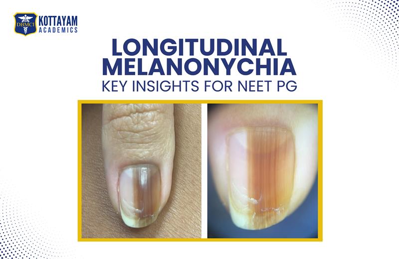
Ever seen a black-to-brown pigmented line going top to bottom on a nail? The pigmented line is Longitudinal Melanonychia (LM). This could be a sign of benign or even a subungual melanoma, a life-threatening condition.
A NEET PG aspirant must know LM in detail, as this comes in the image-based and one-liner set of questions under the subject of Dermatology and Venereology.
Scrolling down, you will find a relevant case presentation, differential diagnosis, investigations, and other necessary information to learn about LM.
Differential Diagnosis
The causes of Longitudinal Melanonychia are of two types. Benign and malignant.
Benign Causes:
- Ethnic Melanonychia – This issue is commonly seen in individuals with darker skin tones. The increase in melanin production in the nail matrix is the cause. A uniform single pigmented line on the nails is an indication.
- Trauma or Friction – It is commonly seen in people wearing tight shoes or having repetitive nail trauma. The stimulating factors are constant rubbing, nail biting, or injury. These will lead to local pigmentation or linear dark streaks.
- Drug-Induced Melanonychia – People taking zidovudine, minocycline, hydroxyurea, and cyclophosphamide can have this issue of nail pigmentation.
- Benign Melanocytic Hyperplasia – This issue occurs due to an increase in the melanocytes in the nail matrix. The increase produces a consistent and uniform pigmentation band. Hyperplasia is a harmless and stable variant.
- Nail Matrix Nevus – This is found in younger individuals. It appears as a single, well-defined, pigmented band. It develops as a benign mole or nevus. The band stays for years.
Malignant Cause:
Subungual Melanoma is a skin cancer that is diagnosed under the nail plate, typically in the nail matrix. When a patient comes in with dark streaks or pigmentation on the nail, investigate in detail because it could be Longitudinal Melanonychia. A common indicator is age. Adults above 50 are commonly found to face this issue.
The pigmented band could also indicate Hutchinson’s sign. It starts from the nail fold or cuticle and extends downward to the nail end. This is an indication of melanoma. Different shades and irregular borders are warning signs of malignancy. A sudden or quickly developing pigmented band on the nail should be diagnosed seriously. Nail deformity, thickening, splitting, and instant bleeding from the affected area are some symptoms of subungual melanoma.
Mistakes to Avoid while preparing for INI CET Exam
As you come closer to the D-day, your exam preparations will tempt you to learn new subjects and facts. Do not fall for it. It only makes your learning complicated and confusing. Learning new topics reduces your overall confidence, while thorough knowledge and understanding of the topics you have already learnt will only make you more confident.
Time management is important in any exam preparation. The time you allocate for theory, practicals, and revision is crucial. Based on the gravity of the topic, spend time wisely or else there are chances of your preparation becoming incomplete. Lack of time management can lead to unnecessary stress.
Avoid comparison. Each individual’s IQ, thought process, aspirations, and goals are entirely different. There is no space for comparison in exam preparation. Each student’s learning process is tailored according to their strengths and weaknesses. Therefore, unnecessary comparison is directly proportional to unnecessary tension and stress.
Over-surfing multiple websites and social media pages for multiple study materials is a common sight. Just because it’s common, it does not mean it is right. Too many resources can create confusion, as different materials can give different information on a single topic. Search for a trusted, clear and concise resource with accurate information and stick to that.
Mnemonic for Red Flags: “ABCDE” of LM
Five factors of Longitudinal Melanonychia are seen as red flags that indicate subungual melanoma. They are:
Age – Adults above 50 have a high chance of pigmentation as the malignant cells increase.
Bandwidth – If the band has a width of more than 3mm, it should be consulted with a doctor.
Change Over Time – Pigmented or irregular bands indicate malignancy and need consultation.
Digit Involvement – The fingers commonly affected are the thumb or great toe. Dark bands on these fingers should be consulted at the earliest.
Extension (Hutchison’s Sign) – If it is a Hutchinson’s sign, the pigmentation vertically starts from the surrounding skin or cuticle to the nail end.
Investigations
Three procedures are conducted to diagnose LM. They are Dermoscopy, Nail Matrix Biopsy, and Histopathology.
Dermoscopy is a safe diagnostic tool used to visualise the pigment patterns in the nail. The parallel longitudinal lines show a benign condition. The irregular, broken, or varying-width lines show melanoma or other malignancies.
Nail Matrix Biopsy is considered the standard for diagnosis. In this process, a small tissue sample is taken from the nail’s growth area, which is the nail matrix. The result will either confirm or rule out melanoma or other causes of pigmentation.
In Histopathology, a biopsy sample is taken to check the melanocyte density, cellular atypia, and architectural disorder. This process shows whether the lesion is benign, dysplastic, or malignant.
Diagnosis
Based on the clinical findings and biopsy results, the most likely diagnosis is a Junctional Melanocytic Nevus of the nail matrix, which presents as benign longitudinal melanonychia (a pigmented band running along the length of the nail).
Four features prove that it is a Junctional Melanocytic Nevus. They are:
- Onset in childhood – The early-onset pigmentation is a strong indication of Benign Melanocytic Lesion.
- Stable and slow changes – Benignity shows gradual darkening, nail plate destruction, or periungual pigmentation.
- Biopsy findings – Normal melanocytes with no atypia or abnormal proliferation, and melanocytes located at the dermoepidermal junction are the findings. Normal melanocytes rule out melanoma, while melanocytes are a characteristic of a junctional nevus.
- Clinical patterns – The pattern found is an evenly pigmented brown band and cracks from a minor trauma.
The final impression is Benign Junctional Melanocytic Nevus of the Nail Matrix presenting as Longitudinal Melanonychia. It is a non-cancerous condition. Periodic monitoring is essential to find any future changes in the pigmentation pattern or nail morphology.
Management
The real challenge for nail experts remains whether a benign lesion appearing in childhood can become a malignant lesion in adulthood. Dermoscopic patterns that suggest a melanoma in children can also be seen in LM, and their specificity in a young age is very low; a brown background with longitudinal brown-to-black lines, with an irregular degree of color pigmentation, spacing, or varying thickness and ending abruptly, or a parallelism disruption, can be observed.
We recommend the examination of all aspects of melanonychia in children when involving a single digit. In our experience, the clinical features of melanonychia in children are the same as in adults, but the most important difference is the extreme variability of the dimensions, degree of pigmentation, and distribution of the pigment, which evolve differently over time. All these characteristics should not cause concern for the clinician when the patient is a child.
Quick MCQ Sample
Q: A 55-year-old patient presents a single dark pigmented band on the thumbnail, 5 mm wide, with pigment extending onto the cuticle. What is the most likely diagnosis?
- Ethnic melanonychia
- Nail matrix nevus
- Subungual melanoma
- Drug-induced melanonychia
Explanation: The pigment spreading onto the cuticle indicates Hutchinson’s sign. The bandwidth being more than 3mm and the age are classic indicators of subungual melanoma. Ethnic or drug-induced melanonychia is ruled out as it usually affects multiple nails and shows uniform, narrow bands. Nail Matrix Nevus is also ruled out as it typically occurs in younger individuals and remains stable over time.
Conclusion
Longitudinal Melanonychia is a sign. A warning that comes in different variants and connects to major types of skin cancers. Knowing about this is important on a general basis. But, as a NEET PG aspirant, this knowledge is highly applicable professionally and personally. Whatever you learn now will come as questions and options during your exam. So, focus on every little detail you find and learn it at the exam level. Therefore, your detailed knowledge about LM is not just a ticket for passing NEET PG, but an early cure for someone going through the disease unknowingly due to some misconceptions.